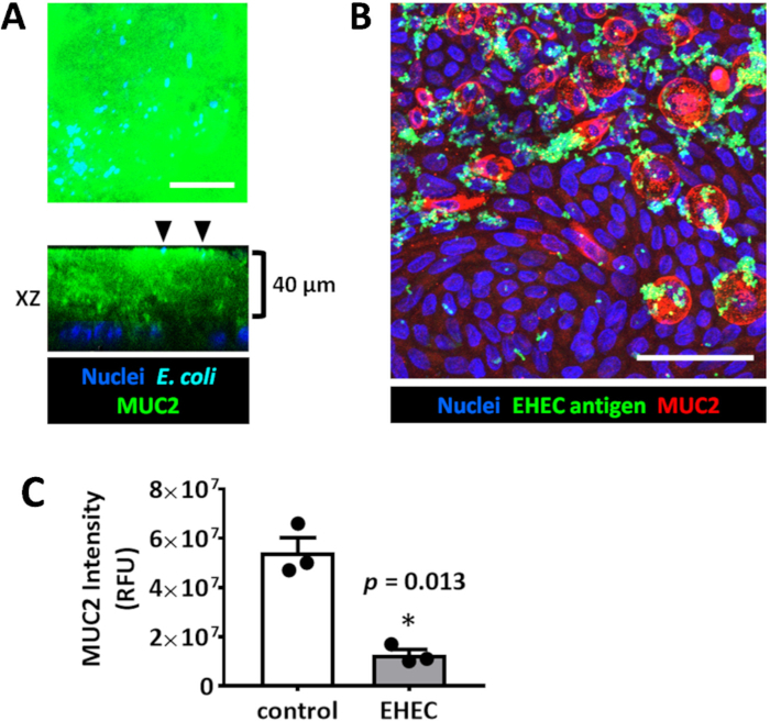
Figure 4: Enteroid/colonoid monolayers are suitable models to study luminal microbe-host interactions.
(A) Representative maximum intensity projection and orthogonal optical section of a colonoid monolayer shows the thick apical MUC2-positive mucus layer which is not permeable to E. coli HS (black arrowheads) sitting at the apical mucus surface. The monolayer was infected for 6 hours with 106 cfu/mL HS. (B) Representative maximum intensity projection of human colonoid monolayer apically infected with EHEC (106 cfu/mL, 6 hours). Scale bar (A–B) = 50 μm. (C) MUC2 immunofluorescence intensity measurements show a significant decrease in MUC2 in EHEC-infected colonoid monolayers compared to uninfected controls. Error bars represent SEM.

