
Fig. 4.
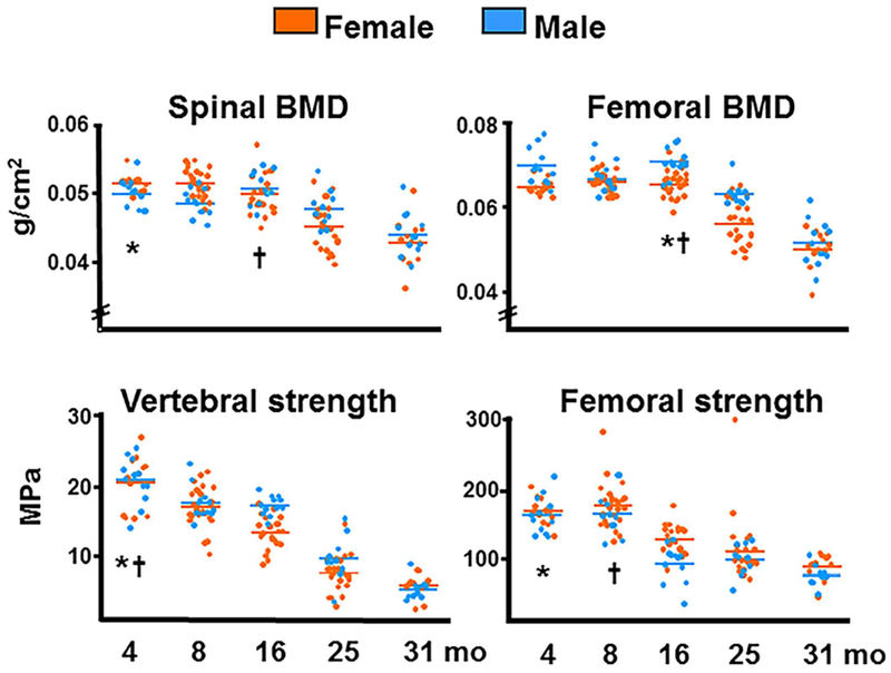
BMD and bone strength decrease with age in C57BL6/J mice. Spinal and femoral BMD was assessed by dual-energy X-ray absorptiometry; strength, by compression testing of the sixth lumbar vertebra and by three-point bending of the left femur. Colored horizontal lines indicate the mean values for each sex. * and † indicate the age after which a time-dependent decline began in females and males, respectively. Reproduced with permission from Almeida and colleagues.(21)