
Fig. 7.
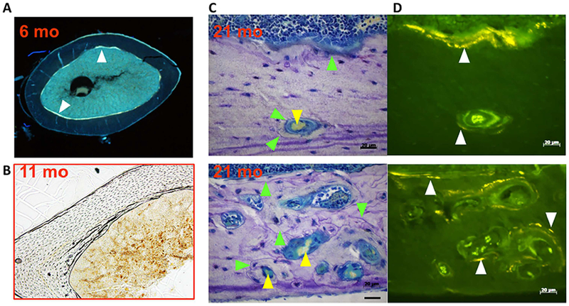
Cortical porosity results from de novo intracortical remodeling. (A, B) Cross-sections from the femoral diaphysis of 6-month-old and 11-month-old C57BL6/J mice. (C, D) Sequential sagittal sections of the femur of 21-month-old mice; the upper and lower panels are from two different sites. Toluidine blue–stained bright field images (C); and unstained adjacent section under fluorescence illumination (D), to visualize tetracycline given at 7 and 3 days before euthanasia. White arrowheads mark tetracycline labeling, green arrowheads mark cement lines, and yellow arrowhead mark red blood cells. In A, the endosteal surface is labeled by tetracycline. Please note lack of intracortical remodeling in B. Please note ample endocortical remodeling as indicated by scalloped cement lines in C and fluorochrome labeling in D. Cement lines and fluorochrome labeling form boundaries around both small and large pores containing a central capillary. Cement lines and tetracycline labels are similarly seen at the endosteal surface in C and D. Reproduced with permission from Piemontese and colleagues.(22)