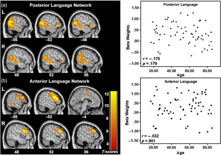
Fig. 2.
Two networks comprising posterior (a) and anterior (b) aspects of typical language brain networks and their corresponding scatterplots of the network’s beta weights with participant age. No significant differences in language network expression with age are observed. Select slices are displayed for each network. The data are shown FWE corrected at p < .001 with 20-voxel cluster threshold. MNI Z-coordinates are displayed beneath each slice.

