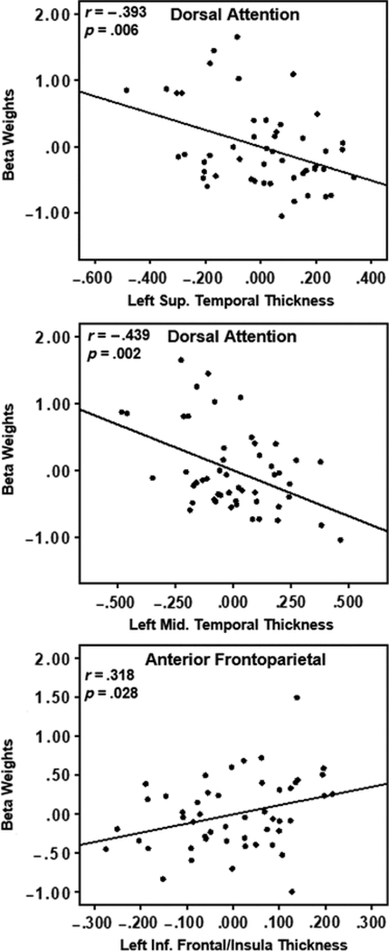
Fig. 4.
Partial correlation scatterplots of average gray matter thickness and network beta weights after controlling for participant age and gender. The top two plots show that increasing atrophy in left temporal regions is correlated with increased recruitment of the anterior aspect of the FPN. The bottom plot shows that increasing atrophy in a left inferior frontal region is correlated with decreased recruitment of the posterior aspect of the FPN.

