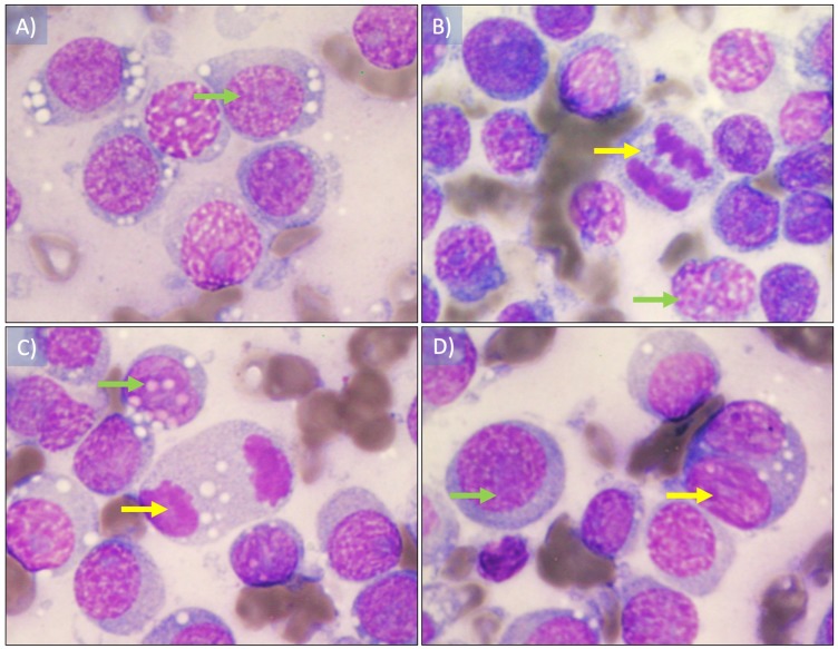
Figure 2. Cytological imprint of canine transmissible venereal tumor.
(A) Red blood cells as well as round to ovoid cells with vacuolated eosinophilic cytoplasm are observed, also hyperchromatic round nucleus with one or two conspicuous nucleoli (green arrow). (B and D) mitotic figures observed in early and (C) later anaphase (yellow arrows). The plasmocitoid morphology tumor cells was 57% (continuous blue arrow) and the lymphocytoid morphology was 43% (blue dashed arrow), the nucleus-cytoplasm ratio was 4:1. Wright staining was used. Images were taken under a 100× magnification.

