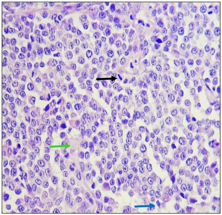
Figure 3. Histological analysis of biopsies.
Tumoral tissue is observed composed of cells that are arranged in mantles and in a dispersed manner, which have scanty, ill-defined eosinophilic cytoplasm and oval nuclei with fine granular chromatin and conspicuous nucleoli, identifying mitosis in a dispersed pattern in metaphase (black arrow) and anaphase (blue arrow) mainly. The observed stroma is lax (green arrow) and there are congestive vessels with neoplastic cells on the wall. Hematoxilin and Eosin staining was used. Images were taken under a 40× magnification.

