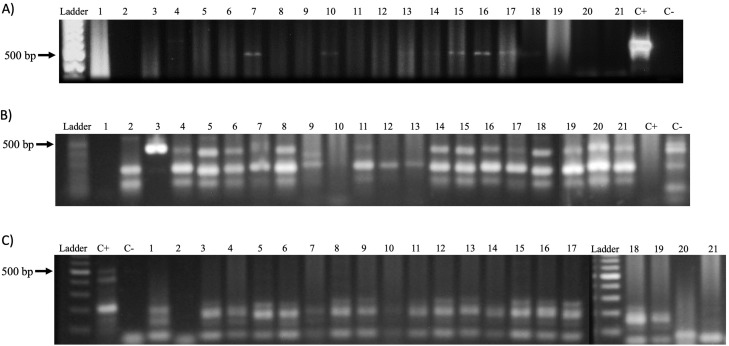
Figure 5. PV detection in samples.
(A) Amplification of the Papillomavirus L1 gene with primers MY09 and MY11 from genomic DNA obtained from biopsies of CTVT. Ladder: Molecular Weight Marker, GeneRuler; lanes 1–21: biopsy samples; H2O: negative control and C+: HeLa DNA (positive control). Samples 7, 10, 13 and 14–17 a band corresponding to the amplicon of the L1 gene of Papillomavirus (450 bp) is observed. (B) Amplification of the L1 gene of Canine Papillomavirus with the primers PVF/FAP-64 from genomic DNA obtained from biopsies of CTVT. Ladder: Molecular Weight Marker, GeneRuler; H2O: negative control and C+: HeLa DNA (positive control); lane 1–21: biopsy samples. A total of 16 of the samples were positive for PV sequences. (C) Amplification of the Canine Papillomavirus E1 gene with the CP4 and CP5 primers. Ladder: Molecular Weight Marker, GeneRuler; H2O: negative control and C+: HeLa DNA (positive control); lane 1–21: biopsy samples. None of the analyzed biopsies was positive.

