Figure 2.

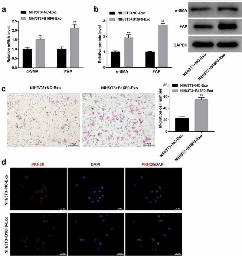
Melanoma-derived exosomes transform fibroblasts into CAFs.
NIH/3T3 cells were co-cultured with exosomes (20 μg/ml) extracted from mouse melanocytes (NC-Exo) and B16F0 cells (B16F0-Exo) for 24 h. (a, b) qRT-PCR and western blot were performed to examine the mRNA and protein levels of α-SMA and FAP, respectively. (c) Transwell migration assay was conducted to evaluate the migration ability of NIH/3T3 cells. Scale bar, 50 μm. (d) Confocal microscope images showed the uptake of PKH26-labeled exosomes in NIH/3T3 cells. PKH26: PKH26-labeled exosomes (red), DAPI: cell nuclei (blue). Scale bar, 25 μm. Values are represented as means ± SD (n = 3). **P< 0.01, vs. NIH/3T3+ NC-Exo.
