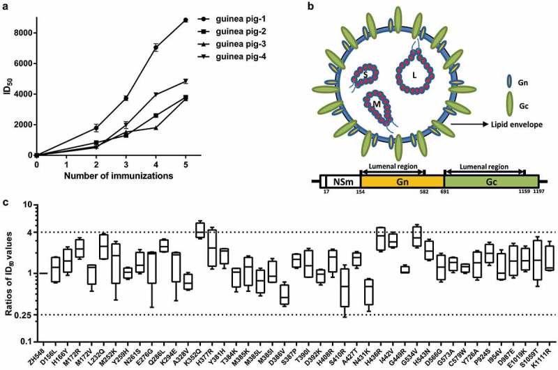
Figure 3.

(a) Relationship between the serum titers from the guinea pigs and the number of immunizations they received. From the second immunization to the last, blood samples were taken from the heart (4 times). (b) Schematic diagram of the structure of RVFV and its M segment. Sites outside the viral membrane are from positions 154 to 582 and from 691 to 1159 (http://www.uniprot.org). (c) The ratios of the ID50 values detected using the pseudoviral variants compared with pRVFV-ZH548. Increases and decreases (4-fold) were highlighted with dashed lines.
