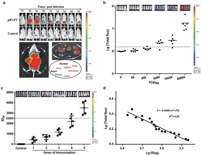
Figure 4.

Establishment of the Balb/c mouse model and determination of neutralizing antibodies in vivo. (a) Determining the optimal detection time point post infection and the targeted organs using BLI. The mouse injected with equal volumes of culture media from the cells transfected with pcDNA3.1+ was used as the control. (b) Determination of the optimal infection dose of pRVFV in vivo. The sum of the mean values (and standard deviations) of the total flux values for the mice challenged with 400 TCID50 of pRVFV was set as the cutoff value to determine a positive or negative result, as highlighted here by dashed lines. (c) Evaluations of neutralizing antibodies in vivo and in vitro. Bioluminescence in the mice was detected by BLI at 6 h pi, and a small serum sample collected from them concurrently was detected in vitro. 2500, the lowest ID50 value for a neutralizing antibody protecting mice from pRVFV infection, as highlighted with dashed lines. (d) Correlation between the total flux values of the mice and the ID50 values of their sera.
