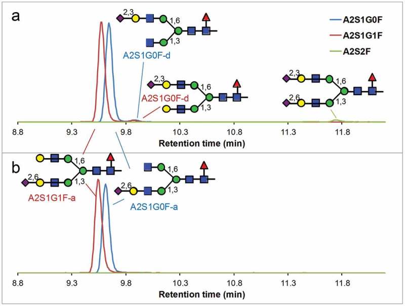
Figure 4.

Reversed-phase profile of sialylated glycopeptides after a 3-h tryptic digestion of human-derived IgG1under a native-like condition (a) and followed by sialidase α-(2–3) treatment (b). Disappearance of the minor peaks (A2S1G0F-d, A2S1G1F-d and A2S2F) indicates that all of these minor peaks contain α2,3-linked sialic acid. The vertical axes represent the relative signal intensities of each SIC.
