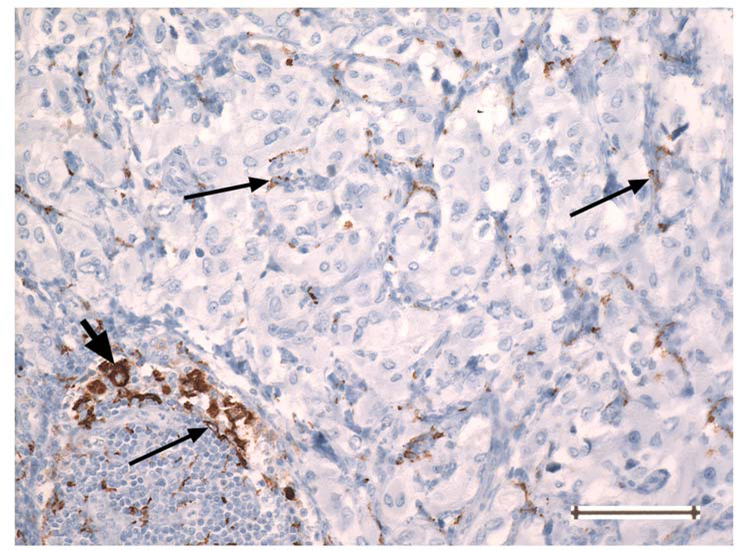
Fig. 4.
PGL showing a CD163-positive macrophage (thick arrow, at left) in an uncommon area of lymphocytic inflammation. The cell size and nuclear size of the macrophage are greater than those of the small CD163-positive monocytes with slender processes (thin arrows) present throughout the tumor. Original magnification 200 ×. Bar =100 um

