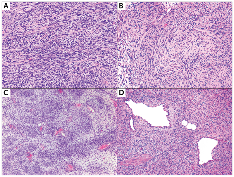
FIGURE 2: Selected histologic features evaluated in the study.

A: Malignant peripheral nerve sheath tumor with intersecting fascicles; B: Cellular schwannoma demonstrating numerous whorls. These whorls are strongly S100-positive (not shown) and named as “Schwannian”; C: Malignant peripheral nerve sheath tumor exhibiting perivascular hypercellularity; D: Malignant peripheral nerve sheath tumor shows nodules of tumor cells herniating into vascular lumens.
