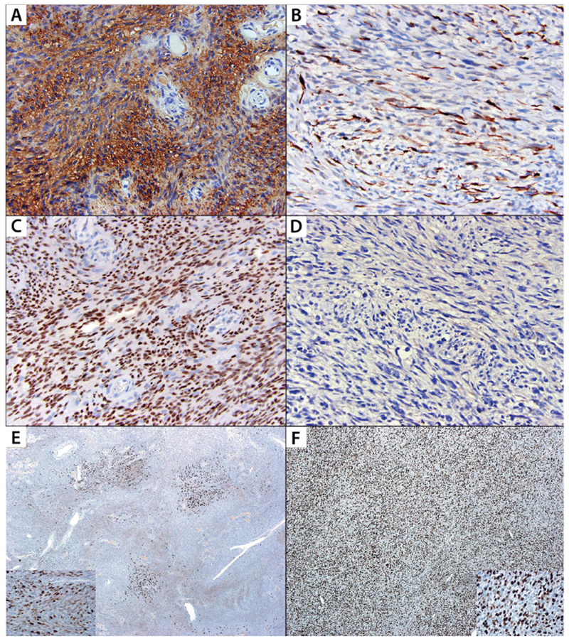
FIGURE 3: Immunohistochemical expression patterns of S100, SOX10 and Ki67 among malignant peripheral nerve sheath tumors and cellular schwannomas.

A: Diffuse S100 positivity in cellular schwannoma; B: S100 highlights occasional cells within a malignant peripheral nerve sheath tumor, which are potentially entrapped Schwann cells; C: Diffuse SOX10 expression in cellular schwannoma; D: SOX10 is negative in malignant peripheral nerve sheath tumor; E: Ki67 stain demonstrates variable labeling indices throughout a cellular schwannoma with hot spots; Inset shows high-power image of a hot spot with labeling index of 36%; F: Ki67 stain demonstrates diffusely increased labeling in a malignant peripheral nerve sheath tumor; Inset shows high-power image with a labeling index of 39%.
