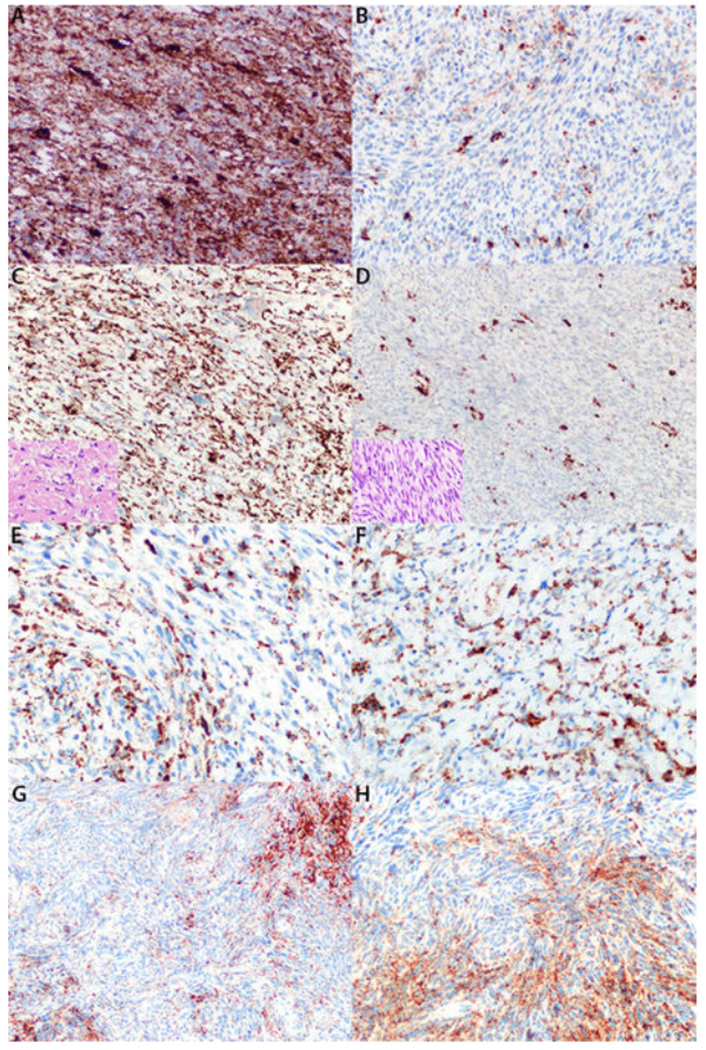
FIGURE 5: Patterns of Patchy Neurofibromin Expression.

A, B: High-grade Neurofibromatosis I (NF1)-associated malignant peripheral nerve sheath tumor demonstrating focal areas of diffuse neurofibromin expression (A) and other areas of neurofibromin loss (B). C, D: NF-1 associated malignant peripheral nerve sheath tumor with low-grade component diffusely positive for neurofibromin (C) and high-grade component completely negative for neurofibromin (D). Inset pictures are hematoxylin and eosin stained sections of corresponding components. E: High-grade NF1-associated malignant peripheral nerve sheath tumor with intermixed positive and negative cells on neurofibromin stain. F: Low-grade NF1-associated malignant peripheral nerve sheath tumor with intermixed positive and negative cells on neurofibromin stain.G, H: Cellular schwannoma with intermixed positive and negative cells on neurofibromin stain, moderate (G) and high power (H).
