FIGURE 3.
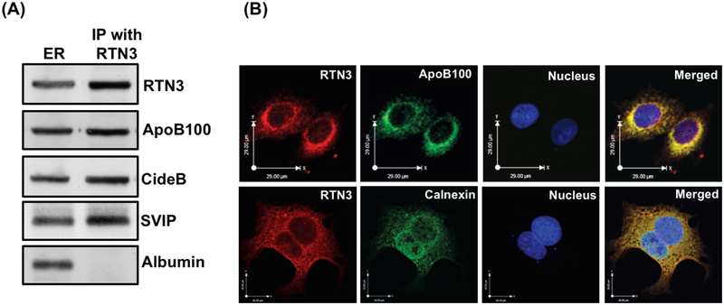
RTN3 interacts with apoB100, cideB and SVIP but not with albumin. A. ER membranes (250 μg protein) were solubilized in 2% (v/v) Triton X-100 and treated with anti-rabbit RTN3 antibodies (10 μg) for 4h at 4°C. Anti-rabbit IgGs bound to agarose beads were added and incubated overnight at 4°C. Immune-complexes bound to agarose beads were isolated and washed 10 times with ice cold PBS to remove unbound proteins. Protein sample was separated by SDS-PAGE (8–16% gel) and probed with anti-RTN3, anti-apoB100, anti-cideB, anti-SVIP and anti-albumin antibodies. B. RTN3 co-localizes with apoB100 in hepatic ER. Hepatocytes were double labeled with either (upper panel) RTN3 (Texas red) and apoB100 (FITC, green) or (lower panel) RTN3 (Texas red) and calnexin, an ER marker (FITC, green. The nucleus is stained with DAPI (blue). Merged figures show colocalization of RTN3 with apoB100 and calnexin.

