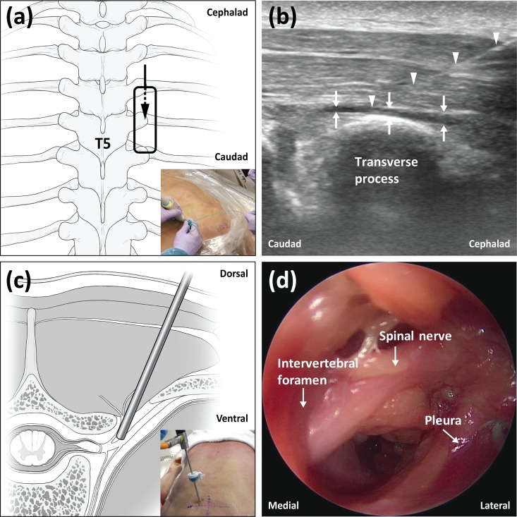
Fig 1. The experimental procedures for injection and endoscopy.
(a) Schematic diagram showing the probe position and needle direction for the erector spinae plane block. (b) Ultrasound image demonstrating needle placement and dye spread of the erector spinae plane block. (c) Schematic diagram representing the endoscope position in the paravertebral space. (d) Typical endoscopic image of the thoracic paravertebral space.

