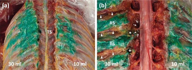
Fig 3. Anatomical dissection findings after erector spinae plane block.
(a) Spread pattern of dye to the fascial layer of the external intercostal muscles after erector spinae plane block with 10 ml (right) and 30 ml (left) of dye. (b) Posterior vertebral bodies were removed. Using an injection of 10 ml (right) of dye, no paravertebral spread was observed. Using 30 ml (left) of dye, T5 and T6 spinal nerves in the intervertebral foraminal area were stained (asterisks), and epidural spread (arrows) was observed at the T5 level. Intercostal nerves were revealed laterally but were not stained (arrowheads).

