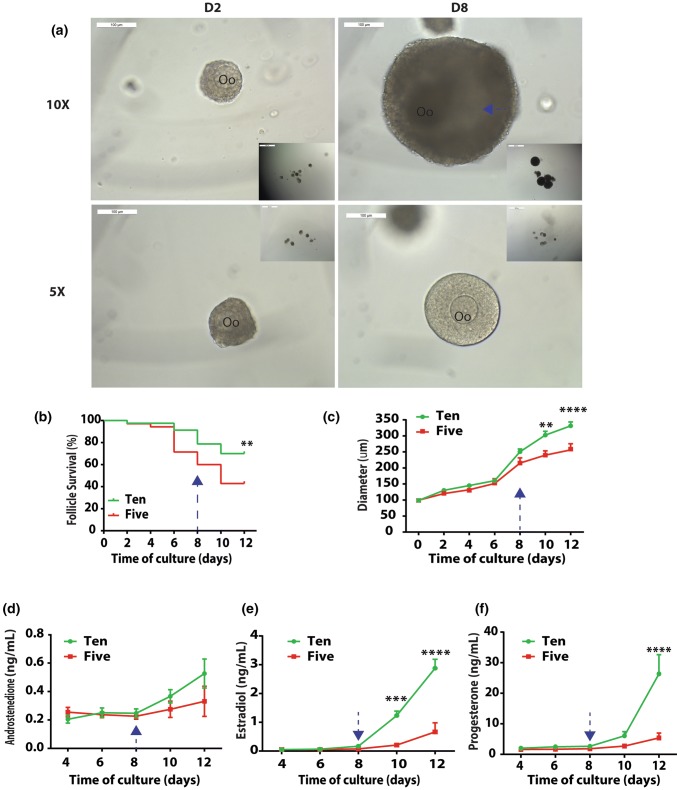
Figure 1.
The number dependent pattern of primary follicle development in vitro. (a) Representative images of 5× and 10× follicles encapsulated in alginate. Scale bar = 100 µm. Oo: oocyte; blue arrow: antrum; (b) Follicle survival in 5× and 10×. Sample size: n = 35 (5×) and 80 (10×); (c) Follicle growth in 5× and 10×. Sample size: n = 15 (5×) and 56 (10×). Data presented as mean ± SEM; Profiles of androstenedione (A4, d), estradiol (E2, e), and progesterone (P4, f) in 5× and 10× follicles during eIVFG. Sample size: n = 4 per time point per condition. Data presented as mean ± SEM, *p < 0.05, **p < 0.01, ***p < 0.001, ****p < 0.0001.

