Figure 3.
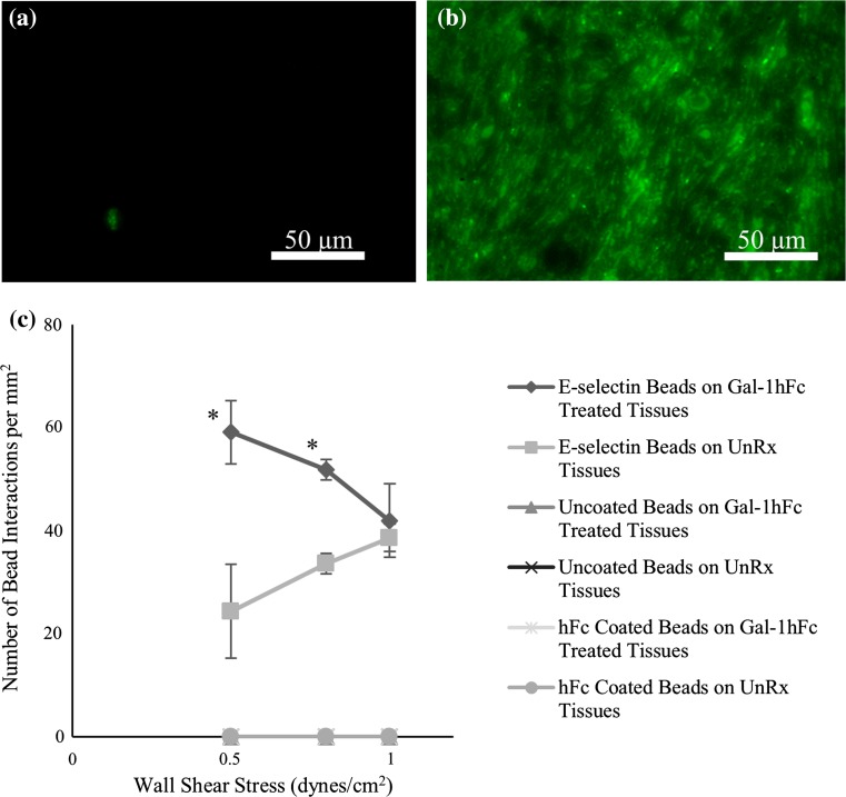
Gal-1hFc treatment of mucinous breast carcinoma tissue microarrays significantly increases E-selectin bead binding at low shear stresses. (a, b) Immunohistochemistry confirms Gal-1hFc binding to mucinous breast carcinoma tissue microarrays. While the untreated control monolayer (a) showed only negligible signal, (b) Gal-1hFc (pseudocolored green) stained quite vibrantly on the treated microarray. Images are representative of n = 3 independent experiments. (c) Analysis of E-selectin-coated, hFc-coated, and uncoated beads’ interactions with untreated and Gal-1hFc treated mucinous breast carcinoma tissue microarrays. Similar to ZR-75-1 breast cancer cells, DBTA of the tissue microarrays exhibited increased binding interactions of E-selectin-coated beads at a wall shear stress of 0.5 dynes/cm2 on Gal-1hFc treated tissues compared to untreated tissues (*p = 0.0014). A significant increase in the number of E-selectin bead interactions between the Gal-1hFc treated and untreated microarrays was also achieved at a wall shear stress of 0.8 dynes/cm2 (* p = 0.0015). Uncoated and hFc-coated beads failed to bind to tissue microarrays under any conditions. Data are mean ± standard error, n = 3 independent experiments.

