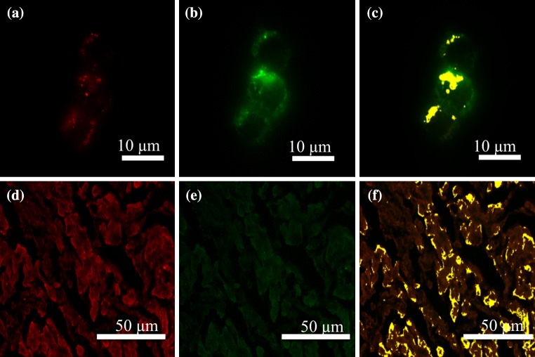
Figure 4.
E-selectin and Gal-1 dual-stained ZR-75-1 breast cancer cells and mucinous breast carcinoma tissues exhibit co-localization of receptor-ligand signals. ZR-75-1 breast cancer cells and mucinous breast carcinoma tissue microarrays were subjected to a dual-staining protocol of murine E-selectin hFc chimera and Gal-1hFc conjugated with Qdot 705. (a) Cells were stained with 20 μg/mL of E-selectin, followed by 10 μg/mL αhIgG AlexaFluor 488 (pseudocolored red). (b) Following E-selectin treatment, Qdot 705 conjugated Gal-1hFc was incubated on the cells at 20 μg/mL (pseudocolored green). (c) Co-localization of E-selectin and Gal-1hFc reactive signals were determined through the inForm 2.0 software and are depicted in yellow. (d, f) Mucinous breast carcinoma tissue microarrays were subjected to a similar dual staining protocol with the exception of αhIgG AlexaFluor 568 used in place of the 488 secondary with E-selectin to reduce autofluorescent background noise from the tissue microarrays. Fluorescent images shown are representative of n = 3 independent experiments.

