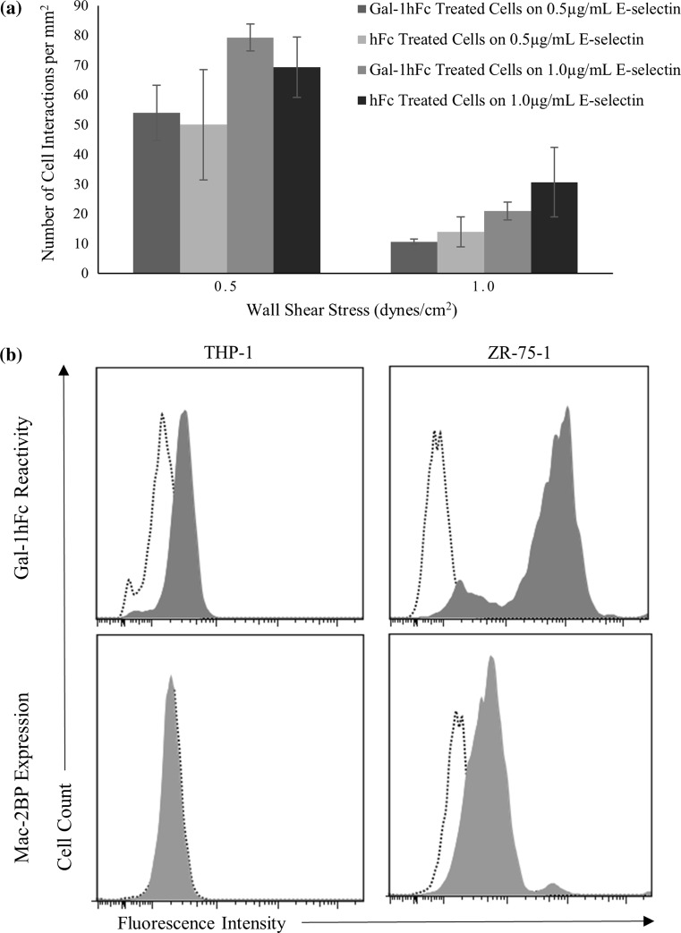
Figure 8.
THP-1 cell adhesion, Gal-1 reactivity and Mac-2BP expression differ as compared to ZR-75-1 cells. (a) THP-1 acute monocytic leukemia cells were treated with 20 μg/mL of Gal-1hFc or isotype control hFc. The cells were then perfused over E-selectin or hFc substrates at a concentration of 0.5 or 1.0 μg/mL, at wall shear stresses of 0.5 and 1.0 dynes/cm2. There were no significant differences (p > 0.05) in interactions between cells treated with Gal-1hFc or with the isotype control. Under any conditions, both Gal-1hFc and hFc cell treatment types had an insignificant amount of binding (2 or fewer events) to hFc coated plates as the negative control. Data are mean ± standard error, n = 3 independent experiments. (b) THP-1 monocytic cells and ZR-75-1 breast cancer cells were labeled with Gal-1hFc (filled) or an hFc control (dotted) (top row) and αMac-2BP pAb (filled) or a rabbitIgG control (dotted) (bottom row), followed by the appropriate secondary antibodies, then analyzed via flow cytometry. ZR-75-1 cells showed relatively high levels of fluorescent intensity for Gal-1hFc and expression levels of Mac-2BP, whereas THP-1 cells had weak to no expression levels of Gal-1hFc reactive sites and Mac-2BP expression levels. Data are representative of n = 3 (Gal-1hFc) and n = 4 (Mac-2BP) independent experiments.

