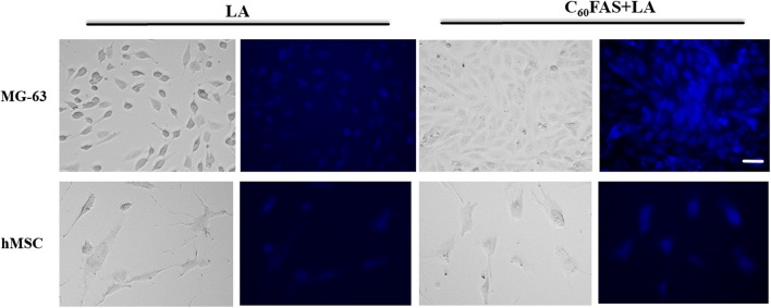
Figure 6.
Transmission light microscopy and fluorescence microscopy of MG-63 and hMSC cells after 2 h of incubation with LA (2.0 μg mL−1) and C60 + LA mixture (1.5 + 2.0 μg mL−1). The blue fluorescence indicates the co-localization of LA and C60 + LA nanocomplex on the cells. Note that C60 fullerene alone is not fluorescent. Scale bar 20 µm.

