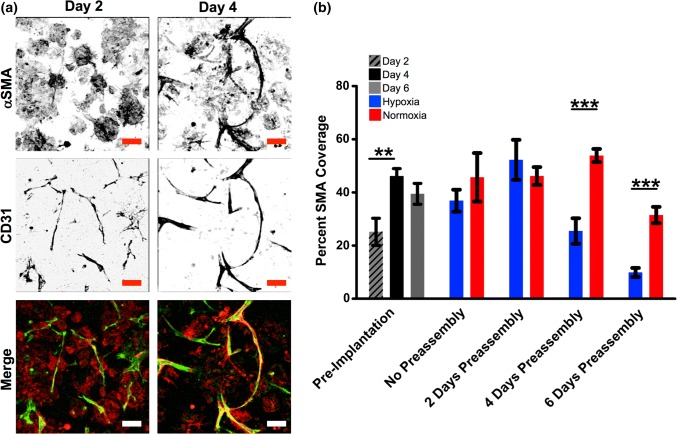
Figure 5.
Analysis of pericyte coverage. (a) Constructs at Days 2 and 4 of preassembly stained for pericytes (αSMA, red) and endothelial cells (CD31, green). (b) Quantification of αSMA+ coverage of CD31+ vessels during vascular assembly of ASCs during preassembly and following transfer to pseudo-implant conditions. **p < 0.005, n = 5. Scale bar = 100 µm.

