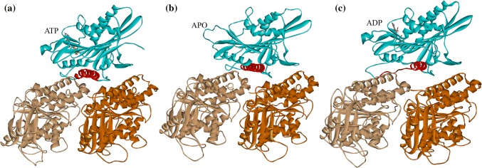
Figure 1.
Stable complex structures of kinesin’s motor domain (blue) with tubulin in different nucleotide-binding states obtained from molecular dynamics simulations. (a) ATP-binding state. (b) apo state. (c) ADP-binding state. Motor domain is shown in blue, α-tubulin in light brown and β-tubulin in dark brown. The binding nucleotides are explicitly shown. α4s of these structures are shown in red.

