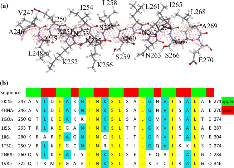
Figure 2.
Residues of kinesin’s α4 helix. (a) Locations of α4’s residues in the helix structure. (b) Sequence alignments of α4s of different kinesin subfamilies (2KIN43 (from Rattus), 4HNA14 (from human) and 1GOJ47 (from N. crassa) belong to kinesin-1 subfamily; 1I5S25 belongs to kinesin-3 subfamily; 1II648 belongs to kinesin-5 subfamily; 1T5C10 belongs to kinesin-7 subfamily; 2NR8 belongs to kinesin-9 subfamily and 1V8J36 belongs to kinesin-13 subfamily). The residues toward motor domain’s core β-sheet (the upper half) and toward tubulin (the lower half) are colored in green and red respectively at the top of the alignments.

