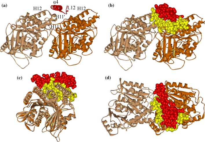
Figure 3.
Binding position of α4 on tubulin. (a) α4 (red helix) locates in the groove between α-tubulin (light brown) and β-tubulin (dark brown) when motor domain (omitted here) binding stably to tubulin. (b) Front view, (c) side view and (d) top view of the geometrical match between α4 and tubulin. The contact atoms (α4 in red and tubulin in yellow) are explicitly shown. The structure used here is the kinesin-1-tubulin complex structure in kinesin-1’s apo state obtained from molecular dynamics simulation.

