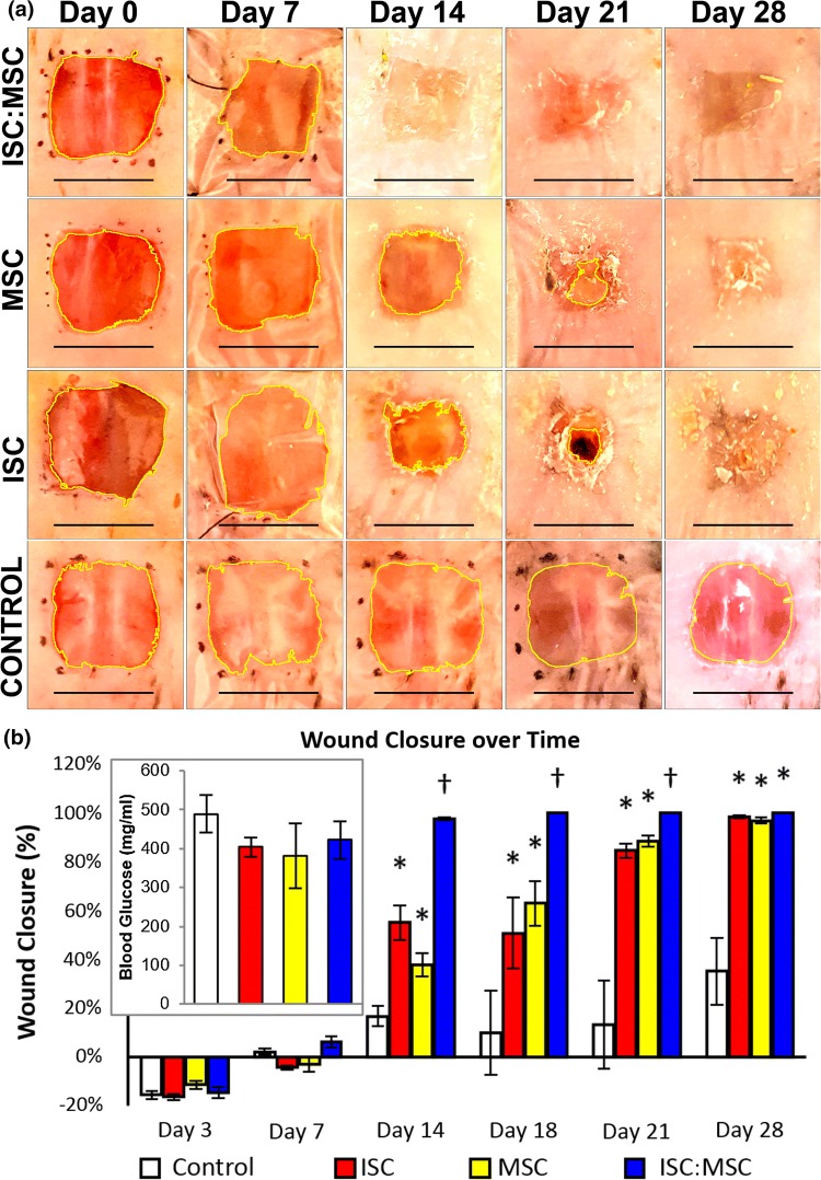
Figure 3.
Progression of wound healing. (a) Rows follow single animals. After Day 0, unhealed wounds are imaged through dressings. Day 14 ISC:MSC shows closed wounds with dried hydrogel residue. Images were uniformly adjusted for contrast, saturation, and brightness. Percent wound closure and glucose levels (inset). Asterisks indicate statistically significant increases (p < 0.05) compared to control; crosses represent significance compared to all other groups. Error bars show standard error of mean.

