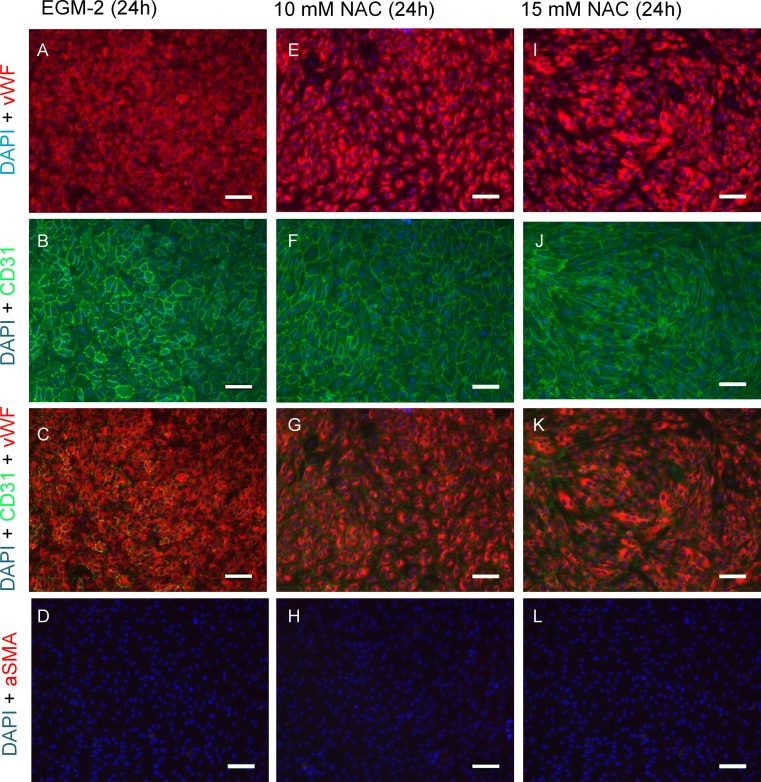
Figure 5.
Immunocytochemical characterization. Confluent HUVEC cell layers after 24 h of NAC supplemented culture. In 15 mM NAC enriched cultivation, endothelial cells changed their morphology from typical cobblestone pattern (a–d) to an elongated, quasi-fusiform configuration (i–l). A slight cellular rearrangement was already detectable at 10 mM (e–h). Cells were positive for common endothelial cell markers CD31 (PECAM-1) and Von-Willebrand-Factor (vWF). Nuclei were counterstained with DAPI. All cells were negative for alpha smooth muscle actin (αSMA), a common myofibroblast marker (scale bar: 100 µm).

