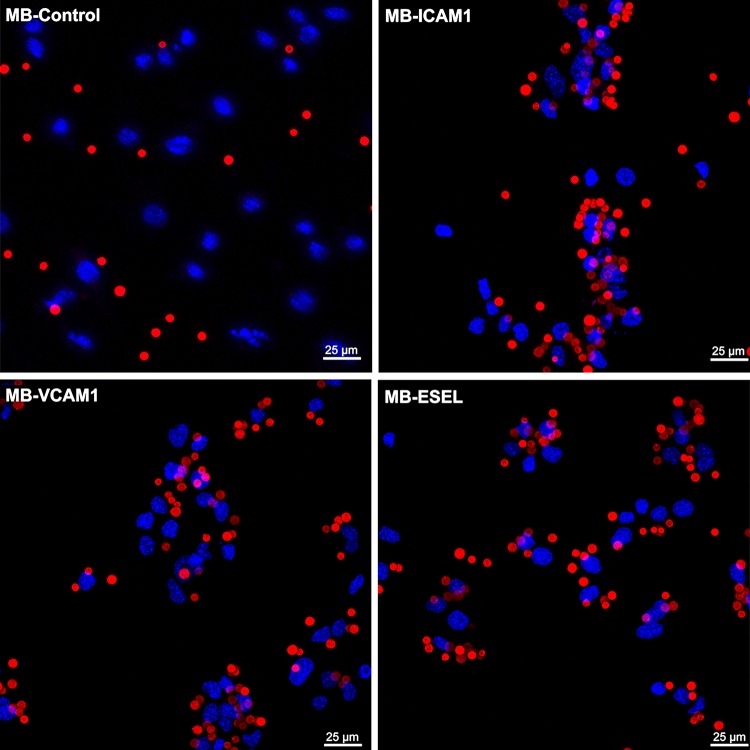
Figure 4.
Maximum-intensity projection confocal laser scanning microscopy images of antibody-labeled MBs adhered to MoAoECs. Images show MBs (Cy3 red); MB-control (MB-streptavidin), anti-ICAM-1-MB, anti-VCAM-1-MB, and anti-E-selectin-MB adhered to endothelial cells (DAPI: blue nuclear staining). Images were acquired by Z-stack (step size 0.99 µm) with a 63× oil immersion objective. ESEL E-selectin.

