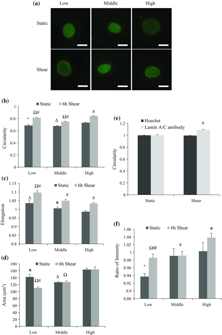
Figure 3.
Shape analysis of endothelial nuclei for groups with different PDL. Cells were labeled with lamin A/C antibody either in static condition or after 6-h shear. At least 28 nuclei were picked for each analysis. (a) Fluorescent images of nuclei stained by anti-lamin A/C antibody before and after shear. Upper panel shows cells in static state, and lower panel shows cells after 6-h shear. Low, middle, and high PDL cells were presented in separate columns, from left to right. Scale bar: 10 µm. (b) Circularity of nuclei with varied PDL before and after shear. All groups of cells had increased circularity by shear stress (# p < 0.001 compared to static), and when compared with high PDL group, low and middle PDL groups all showed significant decrease in either static or shear case (+ p < 0.01, Δ p < 0.001 compared with high PDL group in static state, and Ω p < 0.001 compared with high PDL group after 6-h shear). (c.) Elongation of nuclei with varied PDL before and after shear. All groups of cells had elongated nuclei by shear stress (# p < 0.001 compared to static), and when compared with high PDL group, low PDL group showed significant increase in either static or shear case (Δ p < 0.001 and Ω p < 0.001 compared with high PDL cells in static and shear case, respectively), while middle PDL group only showed significance in control cells compared with high PDL control cells (*p < 0.05). (d) Nuclear area (µm2) of cells with varied PDL before and after shear. Cells with low PDL had decreased area after shear (# p < 0.001), while no significance was observed within either middle or high PDL groups. When compared with the high PDL group, low and middle PDL groups all showed significant area decrease in either static or shear case (*p < 0.05, Δ p < 0.001 compared with high PDL in static state, and Ω p < 0.001 compared with high PDL after 6-h shear). (e) Circularity of nuclei with middle PDL before and after shear stained either by Hoechst solution or anti-lamin A/C antibody. Only nuclei labeled with anti-lamin A/C antibody showed increased circularity after exposed to shear stress (# p < 0.001 compared to static). (f) Ratio of lamin A/C intensity at the nuclear periphery over whole nucleus. Both low and high PDL group showed significant increase in intensity ratio after shear (ϕ p < 0.01 and *p < 0.05, respectively). Significant increase in ratio was also observed in high PDL in static state (+ p < 0.05 compared to low PDL static cells). Moreover, sheared high PDL group showed significant increase compared to the other two (Ω p < 0.001 and π p < 0.05, respectively).

