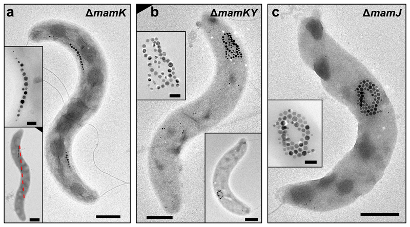
Figure 2. MamY is essential to support the fragmented magnetosome chains in the mamK deletion mutant.
(a) TEM images of ΔmamK cells displaying fragmented magnetosome chains that follow a path along the positive cell curvature (indicated by the red-dashed line - lower inset).
(b) TEM micrographs of the mamY-mamK double deletion mutant, which is unable to organize magnetosomes into chains and displays aggregated magnetosomes.
(c) TEM image of a ΔmamJ cell that presents aggregated magnetosomes. Insets: magnification of the magnetite crystals. TEM images showing corresponding magnetosome configurations were obtained from at least five independent experiments and cell preparations. Scale bars: 500 nm. Insets: 200 nm (except lower inset in A: 500 nm).

