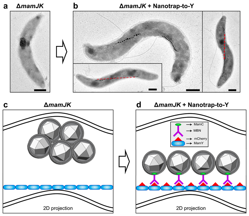
Figure 5. Reconstruction of a synthetic MamY-supported magnetosome chain.
(a) TEM micrograph of the mamJK double deletion mutant displaying clustered magnetosomes.
(b) Several ΔmamJK cells transformed with the Nanotrap-to-Y (MamY-mCherry and MamC-MBN) displaying artificially restored magnetosome chains onto the MamY structure. Red line: projection of the geodetic axis in 2D.
(c) Model of a ΔmamJK cell with clustered magnetosomes localized at the positive curvature. Blue: MamY structure. Note that this is a 2D projection resembling the cells observed by TEM as in (a), therefore, the MamY structure seemingly detaches from the membrane. However, it is continuously associated to the inner positive curvature of the cell.
(d) Model of a ΔmamJK cell plus the Nanotrap-to-Y (mCherry-MamY and MamC-MBN), where the antigen-antibody interaction (mCherry-MBN) allows the artificial recruitment of the magnetosomes to the MamY structural element. Scale bars: 500 nm. Similar magnetosome configurations were observed in cells from three experiments using independent mutant strains and cell preparations.

