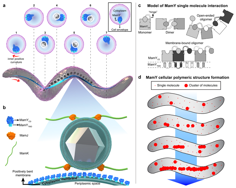
Figure 6. Model for MamY molecular interaction, intracellular polymer formation and localization, and its function as part of the magnetoskeleton.
(a) 3D-rendered illustration of a helical Mgryph cell body (purple) highlighting the localization of the MamY structure (blue) which continuously follows the inner positively-curved membrane. Cross sections depict the position of the MamY structure at different positions along the cell. The MamY structure recruits, scaffolds and organizes the magnetosome chain (membrane-bounded grey crystals) along the geodetic axis.
(b) The magnified model depicts MamY as a transmembrane protein assembling into a continuous structure (blue molecules) attached to the cytoplasmic membrane. An indirect MamJ-MamY interplay is also suggested. This model represents a 2D-projection of a longitudinal section of a helical Mgryph cell, causing the membrane to appear bent. MamY cytoplasmic domain: blue; MamY TM domain: light-blue; MamJ: orange; MamK filament: green; magnetite crystal: grey; magnetosome and cytoplasmic membranes: dark green.
(c) Mechanistic model of MamY monomer interaction by 3D domain swapping. Interactions result in formation of either soluble open-ended oligomers (in the absence of the TM domain) or a stable patch-like layer when anchored to the cytoplasmic membrane (lower scheme) further described in the Supplementary Discussion.
(d) Model for de novo assembly of long-range structures and curvature sensing by MamY. First, MamY oligomers are formed, generating membrane patches large enough to become curvature sensitive. Consequently, MamY oligomers become enriched along the inner positive cell curvature, which promotes further directed interactions thereby generating a geodetically positioned MamY structure. Models are not to scale.

