Figure 2.

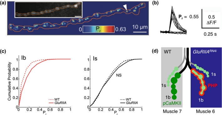
Input and target specificity of PHP. (a) Cumulative AP‐evoked quantal release location heat map derived from postsynaptic Ca2+ imaging at the Drosophila NMJ (SynapGCaMP6f; 200 trials at 0.1 Hz). Inset shows baseline SynapGCaMP6f fluorescence. Local release probability (Pr = number of responses/number of trials at individual sites) is represented as a color scale. Reprinted and adapted from (Newman et al., 2017) with permission from Elsevier. (b) Ca2+ imaging traces (ΔF/F) for the synapse indicated with the arrowhead in (a) during 40 trials. Reprinted and adapted from (Newman et al., 2017) with permission from Elsevier. (c) Cumulative probability for pooled evoked single synapse Pr at wild‐type (WT) and GluRIIASP16 1b NMJs (left) and 1s NMJs (right). Note the increased Pr at type 1b boutons of GluRIIA mutants. Reprinted and adapted from (Newman et al., 2017) with permission from Elsevier. (d) Cartoon illustrating PHP input and target specificity. At the Drosophila NMJ, two motor neurons (“type 1s” and “type 1b” synapses) innervate two muscle cells (“Muscle 6” and “Muscle 7”). PHP (red) is predominantly expressed at type 1b motor neuron boutons contacting the muscle cell with perturbed glutamate receptor function (“GluRIIARNAi”; G‐14‐Gal4 > UAS‐GluRIIARNAi, (Li, Goel, Chen, et al., 2018). This is correlated with reduced phosphorylated CaMKII levels (“pCaMKII,” green; light green indicates reduced pCaMKII levels)
