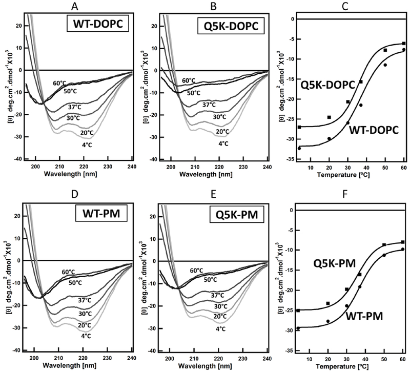
Figure 3. Thermal unfolding profiles of WT and Q5K in liposomes made of DOPC and phagosome-mimic (PM) lipids.

Far-UV CD measurements of the secondary structure of WT and Q5K proteins (100μM) were recorded in 20 mM Tris and 100 mM NaCl pH 7.4 buffer at the indicated temperatures in the presence of 800 μM of the liposomes. The spectra are shown in (A, B, D, E). The mean residue ellipticity at 208 nm (θ208) for both WT and Q5K were plotted as a function of temperature and shown in (C and F).
