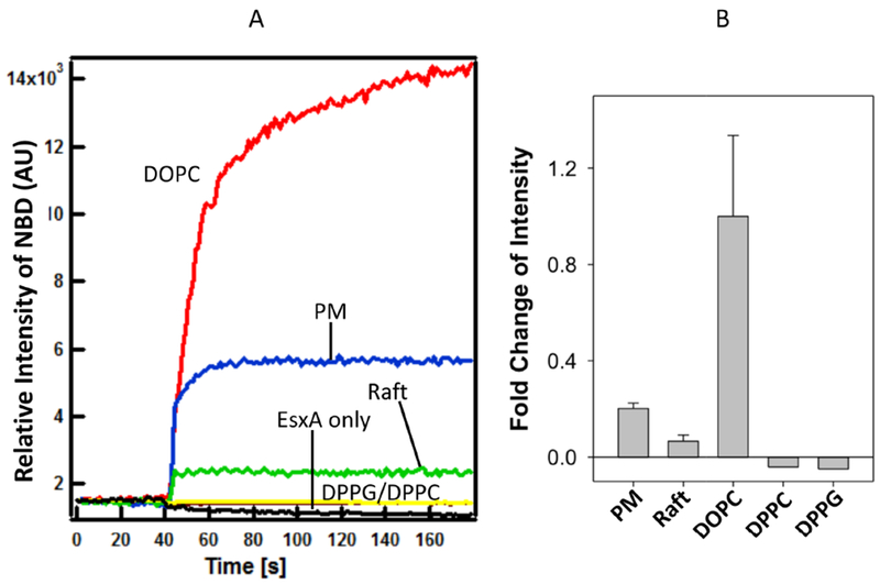
Figure 5. Membrane insertion of EsxA was significantly affected by membrane unsaturation.

The NBD-labeled EsxA WT (3 μM) was incubated with 800 μM of the liposomes containing the indicated lipid compositions (DOPC, DPPG, DPPC, lipid raft, phagosome mimic) at 4 °C for 30 min in 20 mM TrisHCl and 100 mM NaCl pH 7.4 buffer. Then the protein-liposome mixture was transferred at a cuvette at RT. 0.1 volume of 1 M sodium acetate (pH 4) was added to drop the pH. The solution was continuously stirred while recording the data at RT. The emission spectra were recorded at 544 nm with excitation at 488 nm and shown in (A). The fold change of fluorescence intensity relative to DOPC was shown in (B). EsxA without any liposome was used as a negative control. The representative data from 3 experiments are shown.
