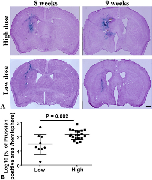
FIG. 3.
Elevation of the AAV-VEGF dose increased bAVM hemorrhage. A: Representative images of Prussian blue–stained sections. The iron depositions (blue) in bAVMs indicate hemorrhage. The nuclei were counterstained with Fast Red. Bar = 1 mm. B: Quantification of Prussian blue–positive area. Log10: the data were 10 log-transformed. Numbers in subgroups were as follows: n = 9 for low-dose AAV-VEGF group and n = 20 for high-dose VEGF group.

