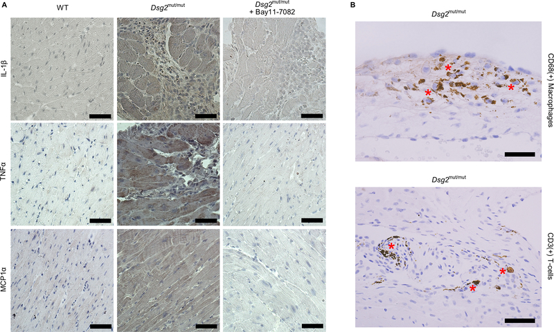
Figure 5. Cytokine expression in cardiac myocytes and infiltrating inflammatory cells in hearts of Dsg2mut/mut mice.
A. Representative immunoperoxidase stained sections of myocardium from vehicle-treated wildtype (WT) mice, Dsg2mut/mut mice and Dsg2mut/mut mice treated with Bay 11–7082 showing immunoreactive signal distributions for IL-1β, TNFα and MCP1α. Signal intensities for all 3 cytokines were increased in myocardial sections from Dsg2mut/mut mice. Signals for IL-1β and TNFα were seen in both cardiac myocytes and infiltrating inflammatory cells in hearts of Dsg2mut/mut mice. Treatment with Bay 11–7082 reduced signal intensity. B. Immunoperoxidase stained sections of myocardium from Dsg2mut/mut mice showing the presence of both macrophages (CD68 + cells) and T-cells (CD3 + cells) (asterisks). Scale bar = 25 μm.

