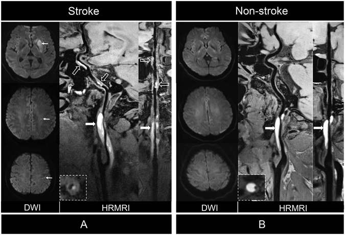
Figure 2.
Comparison of patients with CCAD with or without ischemic stroke. (A) Diffusion weighted imaging (DWI) showed acute ischemic stroke in left basal ganglia and centrum ovale (arrows) of 48 years of male patient. Curved planar reformation of HRMRI demonstrated intramural hematoma (arrows) inner the vessel wall of C1 segment of left internal carotid artery (ICA) and distal intraluminal thrombus (dashed arrows) on the vessel wall of C2 and C4 segments of the ICA. Stretched curved planar reformation HRMRI image depicted irregular surface and intraluminal thrombus. (B) DWI showed no ischemic stroke in a 37 years old female patient. Curved planar reformation of HRMRI image depicted a smooth vessel wall surface (arrows) without intraluminal thrombus. Stretched curved planar reformation of right ICA demonstrated the intramural hematoma and normal distal vessel wall.

