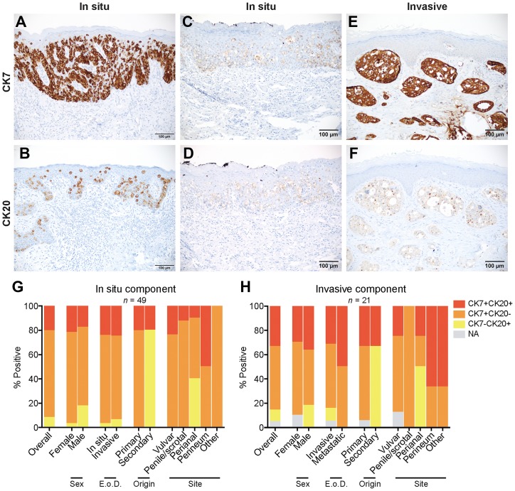
Figure 2. Expression and coexpression of CK7 and CK20 in EMPD.
(A) A CK7 positive/CK20 negative in situ EMPD case shows strong staining intensity (3) in all tumor cells (4). (B) A CK20 positive/CK7 negative in situ EMPD case shows strong staining intensity (3) in all tumor cells (4). (C and D) A CK7 positive/CK20 positive in situ EMPD case shows weak staining intensity (1) in all tumor cells (4) for both markers. (E and F) A CK7 positive/CK20 positive invasive EMPD case shows a strong staining intensity (3) in all tumor cells (4) for CK7 and a weak staining intensity (1) in all tumor cells (4) for CK20. (G) Coexpression of CK7 and CK20 for all tumors with an in situ component (n = 49) categorized by sex, extent of disease (E o D.), origin, and site. (H) Coexpression of CK7 and CK20 for all tumors with an invasive component (n = 21) categorized by sex, extent of disease (E. o. D.), origin, and site.

