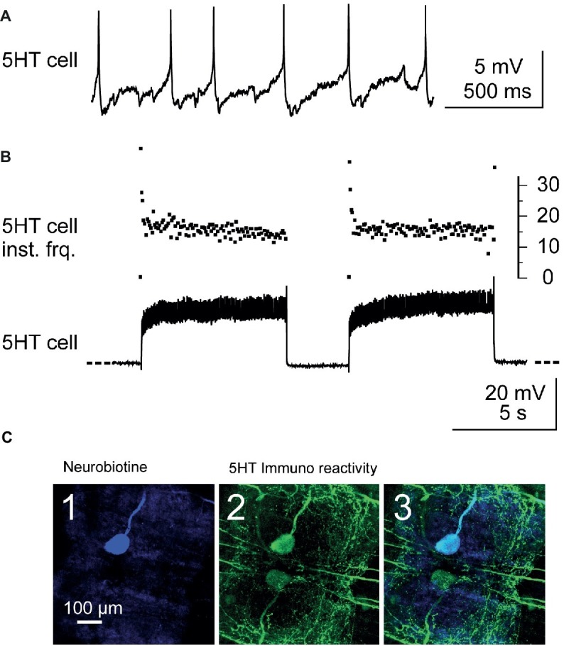
Figure 2.

Identification and stimulation of 5-HT cells. (A) Intracellular recording from a A1 5-HT cell. (B) Stimulation of a 5-HT cell by injection of depolarizing current pulses into the cell body (lower trace). Note the fast adaptation of the instantaneous discharge frequency (5-HT cell inst. Frq.) at the beginning of each current pulse (upper trace): the two first spikes may fire at up to 40 Hz and after the following two spikes, the discharge frequency decreases and stabilizes around 17 Hz. (C) Immunostaining against 5-HT was performed after each experiment in order to confirm that the stimulated neuron was the 5-HT cell. The stimulated neuron was injected with neurobiotin and revealed with Cy5 (ABC kit) and observed in confocal microscopy. The cell body of this neuron appears in blue when illuminated in dark red light (C1). At the same location, a cell body is immunoreactive to 5-HT (green, C2). A superimposition of the two images confirms that the injected neuron is a 5-HT cell (C3).
