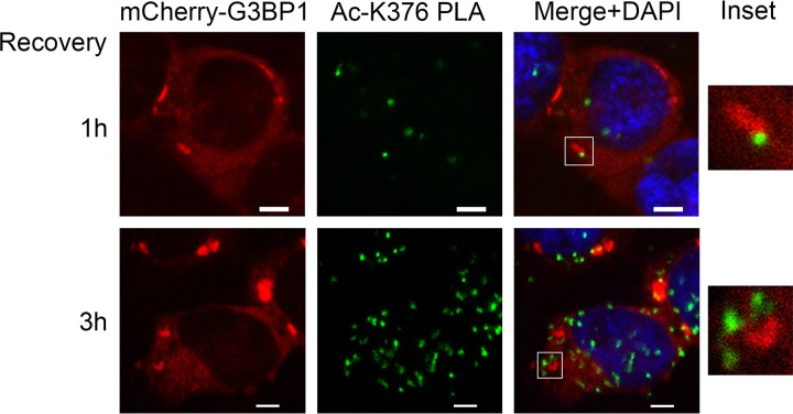
FIG 10.
K376-acetylated G3BP1 localizes primarily outside SGs. Representative confocal microscopic images of mCherry-G3BP1 fluorescence and the K376-acetylated G3BP1 PLA foci (Ac-K376 PLA). The nuclei were visualized with DAPI stain. Acetylated G3BP1/total G3BP1 proximity ligation assay was performed on G3BP1-null 293T-derived cells stably expressing mCherry-WT G3BP1. The cells were pretreated with deacetylase inhibitors for 1 h, followed by SG induction with 0.5 mM sodium arsenite for 1 h and 1 or 3 h of recovery in fresh medium containing deacetylase inhibitors. The acetylated G3BP1 PLA foci were often found in the periphery of the disassembling SGs. The insets show magnifications of the boxed areas. Scale bars, 5 μm.

