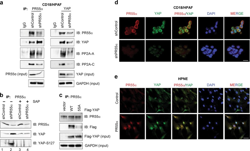
Fig. 6. Interaction of PR55α and YAP in pancreatic malignant and normal cells.
a PR55α was immunoprecipitated from 1 mg protein lysates of CD18/HPAF cells expressing Control-shRNA or PR55α-shRNA with anti-PR55α (100C1) rabbit IgG and probed by immunoblotting for the presence of PR55α, PP2A-C, and PP2A-A subunits with anti-PR55α (2G9), anti-PP2A-C (ID6), and anti-PP2A-A (H300) antibody, respectively. YAP and GAPDH in the lysate were measured by immunoblotting for YAP protein loading control and internal control for protein quantification, respectively. b PR55α was immunoprecipitated from the indicated protein lysates (1 mg per sample) with anti-PR55 (100C1) antibody. The obtained immunoprecipitates were divided into two halves: one half remained untreated (−) and the other half was treated with Shrimp Alkaline Phosphatase (SAP) at 10 units/ml (+) at 37 °C for 1 h. The resulting precipitates were rinsed once with cell lysis buffer and subjected to immunoblotting analysis for PR55α, YAP, and YAP-S127 with the antibody for PR55α (2G9), YAP (D24E4), and YAP-Ser127 phosphorylation (D9W2I), respectively. c PR55α were immunoprecipitated (IP) with anti-PR55α (2G9) antibody from CD18/HPAF cells stably transduced with empty vector, Flag-YAP, or Flag-YAP (5SA) mutant and immunoblotted (IB) using anti-PR55α (100C1) and anti-Flag (M2) antibodies. Lysates from the indicated cells were probed for Flag-YAP (input) and GAPDH (input) with specific antibodies by Western blotting. Intracellular distribution and co-localization of PR55α and YAP in pancreatic malignant and normal cells. The indicated cells were induced for the expression of PR55α-shRNA (CD18/HPAF) (d) or ectopic PR55α (HPNE) (e) by 2 µg/ml Dox for 3 days, and stained with anti-PR55α (100C1) and anti-YAP (1A12) antibodies, as described in the “Materials and methods” section. Images were analyzed for the cellular distribution of PR55α and YAP using a Zeiss-810 confocal laser-scanning microscope. Co-localization of PR55α and YAP in CD18/HPAF cells with/without PR55α-knockdown and in HPNE cells with/without ectopic PR55α expression were examined and shown as merged images (PR55α/YAP and MERGE). Scale bars, 50 µm

