Fig. 4.
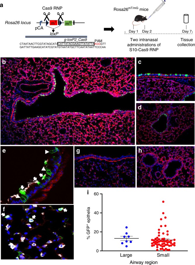
S10 peptide delivery of Cas9 RNP shows editing in ROSAmT/mG locus in vivo. Cas9 RNP directed to loxP sites flanking the tdTomato cassette were administered with S10 peptide once daily on 2 consecutive days. Seven days later, conversion of tdTomato to GFP expression was visualized in lung tissue sections. a Schematic of gRNA targeting two loxP sequences flanking tdTomato gene and experimental protocol for Cas9 RNP delivery to ROSAmT/mG mice. b Fluorescence image of large airway 7 days following two intranasal doses of [S10]: 40 µM; [Cas9]: 1.33 µM; [gRNA]: 2 µM; ×2 magnification. GFP expression denotes edited cells. c Editing in a large airway, ×20 magnification. d Editing in a small airway; ×20 magnification. e Co-localization of GFP and marker of ciliated cells (α-tubulin, white) in large airway. Arrows indicate α-tubulin co-localization with GFP. Arrowheads denote edited (GFP+) non-ciliated cells negative for α-tubulin; ×40 magnification. f Co-localization of GFP+ and SP-C (white) identifies alveolar type II cells (arrows) in distal lung; ×40 magnification. g Representative image of large airway after delivery of Cas9 RNP alone shows no editing, ×20 magnification. h Representative image of small airways after delivery of Cas9 RNP alone shows no editing; ×20 magnification. i Editing efficiency of Cas9 in large and small airways quantified by the number of GFP+ cells. Horizontal lines indicate mean ± SE; n = 5 mice/group. Data underlying this figure are provided as Source Data file

