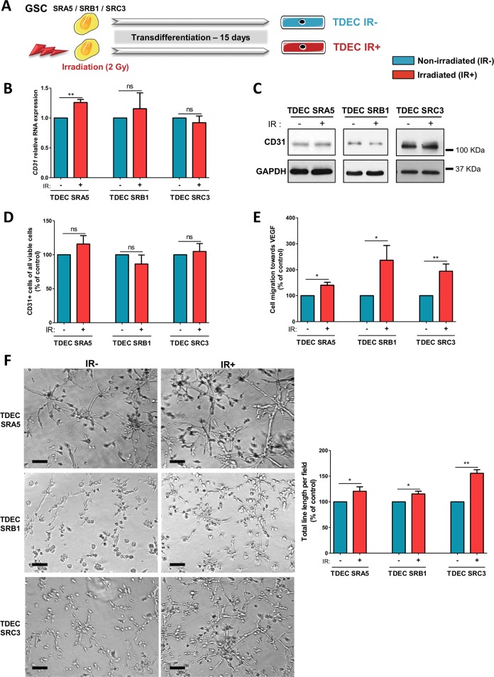
Fig. 2. Ionizing radiation potentiates GSC transdifferentiation into TDEC.
a GSC isolated from tumors of 3 patients (SRA5, SRB1 and SRC3) were or were not irradiated (2 Gy) and were then cultured in EGM-2 for 15 days in order to obtain TDEC IR+ or TDEC IR−. b Relative RNA expression of the endothelial marker CD31 determined by RT-qPCR in TDEC IR− vs. TDEC IR+ from the three different GSC (SRA5, SRB1 and SRC3). The fold inductions are expressed as means ± SEM of at least three independent experiments (normalized to TDEC IR−). c Immunoblot of CD31 in TDEC IR− vs. TDEC IR+. Blots are representative of at least 3 independent experiments in the three patients’ GSC lines (SRA5, SRB1, and SRC3). d Immunofluorescence analysis by FACS of CD31 protein expression in TDEC IR− vs. TDEC IR+. The graphs represent means SEM of the percentage of CD31 positive cells among all viable cells of at least 3 independent experiments. e Cell migration towards VEGF. The graphs represent means ± SEM of the percentage of cells that migrate towards VEGF normalized to TDEC IR−. f Pseudotube formation assay. The graphs represent means ± SEM of the total line length per field determined by the quantification of at least 3 fields per well (normalized to TDEC IR−). Scale bars, 100 µm

