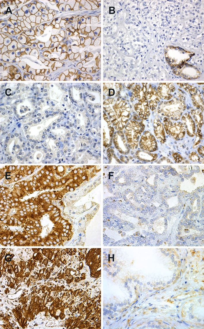
Figure 1.

Immunohistochemical staining of (A–F) localised prostatic carcinomas, (G) castration resistant carcinoma and (H) BPH. (A) Strong membranous E‐cadherin; (B) weak E‐cadherin staining with strong staining in a benign gland at the lower right corner; (C) negative membranous N‐cadherin staining; (D) positive membranous N‐cadherin staining; (E) strong cytoplasmic FOXC2 in carcinoma, weaker staining in the benign epithelium to the right; (F) weak FOXC2 staining; (G) strong cytoplasmic FOXC2 staining in castration resistant carcinoma and (H) weak FOXC2 staining in BPH. Original magnification, ×400.
