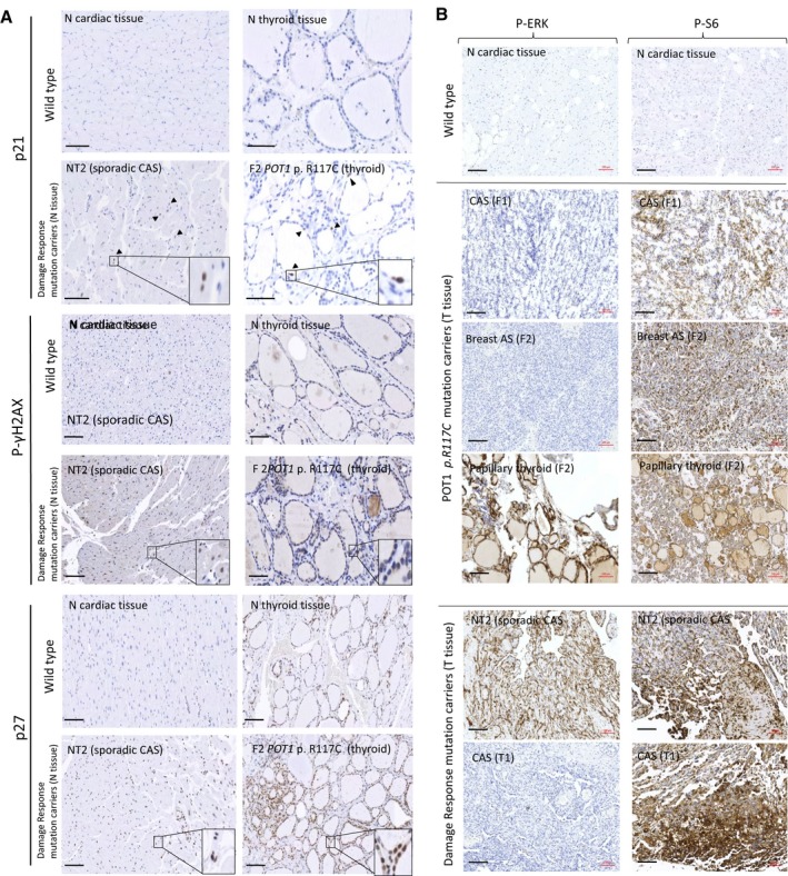
Figure 2.

Immunohistochemical staining. A, Tissue stress was tested in normal tissue of carriers of the POT1 p.Arg117Cys (p.R117C) mutation and carriers of mutations in the damage response‐signaling pathway (sporadic CAS) in comparison with the corresponding normal tissue without mutations (wild type). Above: Wild‐type cardiac and thyroid tissues from healthy controls without mutations. Below: normal tissue of individual NT2 (sporadic CAS individual with constitutional mutations in the ATR,ATM and TP53BP genes) and normal tissue of individual F2 (papillary thyroid tumor with POT1 p.R117C mutation) as representative examples (see Table 2 for all studied individuals). Increased cell cycle arrest was observed in the normal tissue of both patients. Black arrowheads show some of the stained nuclei. Detailed fields (10×) are also shown. Scale bar (in black): 100 μm. B, IHC staining with anti–P‐ERK and anti–P‐S6 antibodies in tumor tissues. Tumor tissues from carriers (F1 and F2) and noncarriers (T1 and NT2) of the POT1 p.R117C mutation compared with a normal tissue section (negative staining) are shown as examples (see Table 2 for all studied individuals). Three tumors from the 2 patients (F1 and F2) carrying the POT1 p.R117C mutation are shown: both angiosarcomas (CAS tumor tissue from F1 and breast AS from patient F2) only showed immunoreactivity with anti–P‐S6 antibody, while the papillary thyroid tumor (patient F2) also showed immunoreactivity with anti–P‐ERK antibody. Two staining patterns were observed in sporadic CAS patients without mutations in the POT1 gene: tissue from patient T1 only showed immunoreactivity with anti–P‐S6, whereas tissue from patient NT2 (sporadic CAS) was stained with both anti–P‐S6 and anti–P‐ERK antibodies. Scale bar (in black): 100 μm. AS indicates angiosarcoma; CAS, cardiac angiosarcoma; N, normal tissue; T, tumor tissue.
