Figure 2.

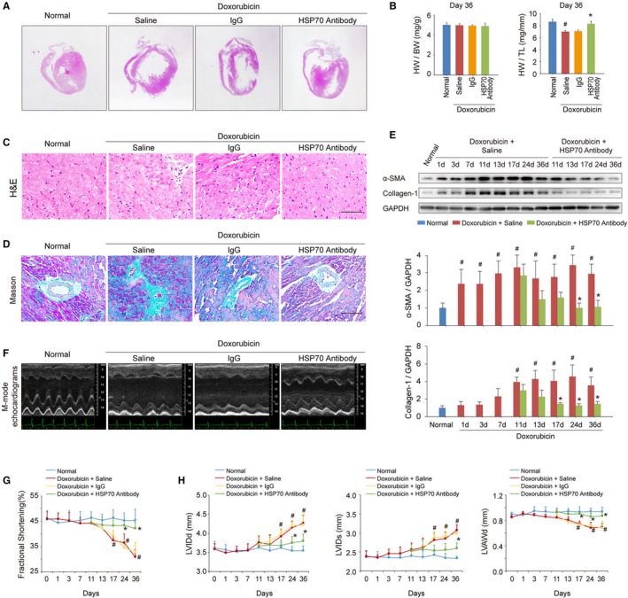
Blocking extracellular HSP (heat shock protein) 70 activity attenuates doxorubicin‐induced cardiac remodeling and dysfunction. A, Blocking extracellular HSP70 with a neutralizing antibody attenuated doxorubicin‐induced ventricular dilatation (global heart section). B, The ratios of heart weight/body weight (HW/BW) and HW/tibia length (HW/TL) are presented (n=6–8/group). # P<0.05, compared with normal mice; *P<0.05, compared with doxorubicin‐treated mice. C, Blocking extracellular HSP70 inhibited the recruitment of inflammatory cells (bar=50 μm), as indicated by hematoxylin‐eosin (H&E) staining of the heart sections. D, Representative photomicrographs of Masson's trichrome staining of the heart sections for cardiac fibrosis evaluation (bar=50 μm). E, Expression of α‐smooth muscle actin (α‐SMA) and collagen I was assayed by Western blot analysis. The expression ratio of the indicated protein to GAPDH from 3 independent experiments is presented. # P<0.05, compared with normal mice; *P<0.05, compared with doxorubicin‐treated mice. F, Representative images of left ventricular (LV) M‐mode echocardiograms (n=6–8/group). Cardiac functional (G) and structural (H) parameters were measured by echocardiography analysis. Data are mean±SEM (n=6–8/group). # P<0.05, compared with normal mice; *P<0.05, compared with doxorubicin‐treated mice. LVAWd indicates LV diastolic anterior wall thickness; LVIDd, end‐diastolic LV internal diameter; LVIDs, end‐systolic LV internal diameter.
